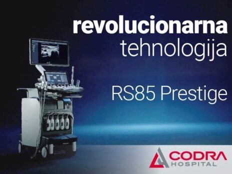
A Novelty in the CODRA Hospital is the ultrasound machine RS85 Prestige – revolutionary technology with new diagnostic characteristics, based on top notch imaging performance.
The latest generation ultrasound device RS85 Prestige is a big step forward in ultrasound and doppler diagnostics of all regions of the body.
Advantages of advanced technologies in diagnostic
The high resolution of the image which which allows the display of the smallest details, using different frequencies of ultrasound imaging and combining them into a single image, enables a detailed display with recognizing even the smallest changes in the structure of tissues and organs. 3D Doppler and a unique display of microcirculation within the changes themselves are of crucial importance in diagnostics and in the further monitoring of therapeutic effects in the treated change.
The integration of images from other modalities, scanners and MRI, with an ultrasound image in real time, enables establishment of a diagnosis and monitoring of changes without repeated scans and magnetic resonance examinations, which ultimately leads to less harmful (radiation and contrast administration) and more comfortable examinations and control of the applied therapies, with undoubtedly more information obtained by comparing the two test modalities than having just one, using the absolute advantages provided by each device individually
The integration of imaging and biopsy, guided by electromagnetic waves, reduces tissue trauma in patients and significantly increases the precision of diagnostics, thus enabling faster and more accurate treatment, which is the ultimate goal of all diagnostic procedures.
By applying the elastogarphy method when diagnosing diseases of breasts, thyroid gland and liver with advanced software and by using AI we enable precise diagnostics and quantification of changes in these organs, at the same time reducing subjective impression of the examiner.
The device has hardware and software capabilities for applying contrast ultrasound, which enables precise, fast and accurate diagnosis of focal changes, without the use of harmful X-rays and contrast agents, as well as more frequent, safer and simpler control of identified changes and conditions.
In urological diagnostics, image fusion with magnetic resonance and electromagnetic biopsy guidance significantly improve the precision of biopsies with less tissue trauma and greater accuracy of findings, which is of key importance for fast and accurate therapy.


